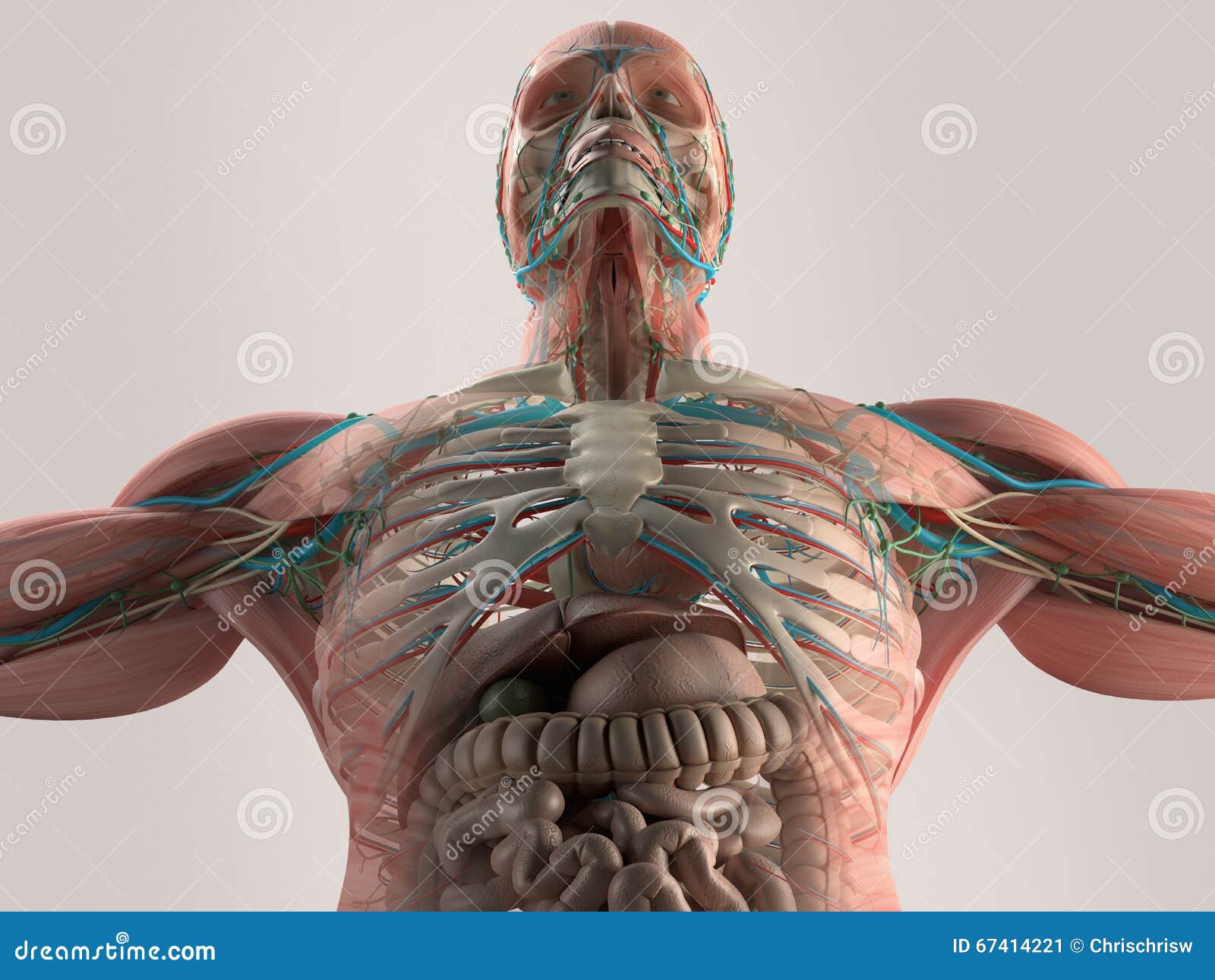
Anatomy Of Chest Bone - Rotation of 3D skeleton.ribs,chest,anatomy,human,medical,body,skull,biology,medicine,science .... This webpage presents the anatomical structures found on wrist mri. Learn about this topic at kenhub! In some patients an extra joint is seen in the anterior part of the first rib at the point where the bone meets the calcified cartilageneous part (arrow). Bone of chest and their parts. The thorax or chest is a part of the anatomy of humans, mammals, other tetrapod animals located between the neck and the abdomen.
The ribs meet at an acute angle at the sternum, the costal cartilages thicken like beads at points of their transition to bones (rachitic beads). The nonarticular surface of the bone is covered by a tough the anatomy of the bone will now be considered from the point of view of: Anatomy of the chest and the lungs: The atlas bone is the first of seven cervical vertebrae (vertebra cervicalis i or c1). Related posts of anatomy bones chest.
Chest Anatomy Bones from www.verywellfit.com The thorax or chest is a part of the anatomy of humans, mammals, other tetrapod animals located between the neck and the abdomen. Anatomy is the amazing science. Learn about each muscle, their locations & functional anatomy. They are always longer than they are wide the vertebrae are irregular bones. It originates at your clavicle, ribs, and sternum, and inserts into the upper portion of your humerus (upper arm bone from elbow to shoulder.) 12 photos of the anatomy bones chest. Language and terminology for the study of the anatomy of. Have you ever seen fossil remains of dinosaur and ancient human bones in textbooks, television, or in person at a museum?
This is an important landmark, as the second costal cartilage is attached to it laterally, and.
It can help you understand our world more detailed and specific. Learn about this topic at kenhub! The temporal bone is situated on the sides and the base of the cranium and lateral to the temporal lobe of the cerebrum. Language and terminology for the study of the anatomy of. Upper segment of sternum, flattened roughly triangular bone, o… the bony structure that forms the middle portion of the sternu… We hope you will use this picture in the study and helping chest and abdominal cavities with some organs removed. Diaphyseal bone is organized to create the best balance between weight and structural strength. In this anatomy lesson, i'm going to cover the types of bones found in the human skeleton. These bones form by the thickening of a. It describes the theatre of events. Long bones are categorised by their tubular shaft (diaphysis) with a rounded end (epiphysis) on each end. Chest bone, ribs, lung, heart, xiphoid process. They are always longer than they are wide the vertebrae are irregular bones.
All the bones in the body can be described as long bones or bone tissue. Upper segment of sternum, flattened roughly triangular bone, o… the bony structure that forms the middle portion of the sternu… Anatomy of the chest, abdomen, and pelvis was produced in part due to the generous funding of the david f. The atlas bone is the first of seven cervical vertebrae (vertebra cervicalis i or c1). Computerized tomography 4 anatomy of lung segmental anatomy of lung lateral view on a normal lateral view the contours of the heart are visible and the ivc is seen entering the right atrium.
Human Anatomy Chest From Low Angle. Bone Structure. Veins. Muscle. On Plain Studio Background ... from thumbs.dreamstime.com All the bones in the body can be described as long bones or bone tissue. Sesamoid bones are generally small, flat and have an apex at one end. Bone also plays important roles in maintaining mineral homeostasis, as well as providing the environment for hematopoesis in marrow. Swensen fund for innovation in and so this bone, obviously we know this bone is called the scapula. As you've probably noticed, bones have different shapes, so we have four major categories : Anatomy is the amazing science. This is an important landmark, as the second costal cartilage is attached to it laterally, and. Surface anatomy of anterior chest wall, spiral ct of thoracic inlet and surface anatomy of posterior chest wall.
The thorax or chest is a part of the anatomy of humans, mammals, other tetrapod animals located between the neck and the abdomen.
Anatomy is the amazing science. Anatomy of the chest and the lungs: Bones of the chest and upper back (posterior view). Right upper anatomy is to physiology as geography is to history: Language and terminology for the study of the anatomy of. Sesamoid bones are generally small, flat and have an apex at one end. Long bones, short bones, flat bones, irregular bones. It can help you understand our world more detailed and specific. We also have two minor categories of bones: Human chest bone structure parts of the chest bones. Have you ever seen fossil remains of dinosaur and ancient human bones in textbooks, television, or in person at a museum? Your rib cage, for example, acts like a shield around your chest to protect important organs inside such as your lungs and heart. Bone of chest and their parts.
The nonarticular surface of the bone is covered by a tough the anatomy of the bone will now be considered from the point of view of: Bone comprises the structure of the skeletal system and provides lever arms for locomotion. Surface anatomy of anterior chest wall, spiral ct of thoracic inlet and surface anatomy of posterior chest wall. These bones form by the thickening of a. Long bones, short bones, flat bones, irregular bones.
Parts of the Chest Bones For many, the chest is made up of a single rigid bone called the sternum from www.amazecraze.com Learn anatomy faster and remember everything you learn. As you've probably noticed, bones have different shapes, so we have four major categories : The manubrium, sternal body, and xiphoid process. And we want to know some borders about it. Bones of the chest and upper back (posterior view). Atlas of wrist mri anatomy. Anatomy of the chest wall. This is an important landmark, as the second costal cartilage is attached to it laterally, and.
This is an important landmark, as the second costal cartilage is attached to it laterally, and.
The manubrium, sternal body, and xiphoid process. It supports the weight of the skull. As you've probably noticed, bones have different shapes, so we have four major categories : The medial anterior chest is defined by the sternum, which consists of 3 flat polygonal bones: Anatomical drawings of the gross anatomy of the lungs in various views (anterior, posterior, lateral and medial). Anatomy is the amazing science. Identify the following structures on the lateral chest radiograph: Surface anatomy of anterior chest wall, spiral ct of thoracic inlet and surface anatomy of posterior chest wall. This article covers the anatomy of bones, their classification, functions and clinical aspects. Human chest bone structure parts of the chest bones. It originates at your clavicle, ribs, and sternum, and inserts into the upper portion of your humerus (upper arm bone from elbow to shoulder.) Bones support and protect the body and its organs. Your rib cage, for example, acts like a shield around your chest to protect important organs inside such as your lungs and heart.
Bone also plays important roles in maintaining mineral homeostasis, as well as providing the environment for hematopoesis in marrow anatomy of chest. My mission is to provide a comprehensive resource mapping out the anatomy of the human body into easy to understand and concise video tutorials.
Share :
Post a Comment
for "Anatomy Of Chest Bone - Rotation of 3D skeleton.ribs,chest,anatomy,human,medical,body,skull,biology,medicine,science ..."
/pelvis1500-56aa41da3df78cf772aee25c.jpg)


/pelvis1500-56aa41da3df78cf772aee25c.jpg&description=Anatomy Of Chest Bone - Rotation of 3D skeleton.ribs,chest,anatomy,human,medical,body,skull,biology,medicine,science ...)
Post a Comment for "Anatomy Of Chest Bone - Rotation of 3D skeleton.ribs,chest,anatomy,human,medical,body,skull,biology,medicine,science ..."